
Mouse Secondary Antibodies


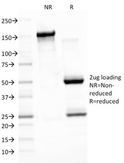
Biotium Epstein-Barr Virus (LMP-1)(CS3), Biotin conjugate, 0.1mg/mL
This antibody is a mixture of four different monoclonal antibodies. This antibody is specific to 60 kDa latent membrane protein (LMP-1) encoded by the BNLF1 gene of the EBV. Each clone reacts with different epitopes on the hydrophilic C-terminus of the cytoplasmic domain of LMP-1. This antibody stains strongly with EBV-positive lymphoblastoid cell lines and EBV infected B cell immunoblasts in infectious mononucleosis. EBV, also designated human herpesvirus 4 (HHV-4), is a member of the herpesvirus family and is one of the most common human viruses. EBV infects B cells and, though often asymptomatic, it can cause infectious mononucleosis, a disease characterized by fatigue, fever, sore throat and muscle soreness. Primary antibodies are available purified, or with a selection of fluorescent CF Dyes and other labels. CF Dyes offer exceptional brightness and photostability. Note: Conjugates of blue fluorescent dyes like CF405S and CF405M are not recommended for detecting low


Biolegend Spark Blue 574 anti-mouse CD90 2 (Thy1 2) [30-H12] Isotype Rat IgG2b Reactivity Mouse Apps FC Size 25 ug
Spark Blue 574 anti-mouse CD90 2 (Thy1 2) [30-H12] Isotype Rat IgG2b Reactivity Mouse Apps FC Size 25 ug

Biotium CD53(63-5A3), CF740 conjugate, 0.1mg/mL
Recognizes a protein of 33-55 kDa, identified as CD53 (Workshop V; Code CD53.1). CD53 is expressed on monocytes, and macrophages, granulocytes, dendritic cells, osteoblasts and osteoclasts, NK cells, and on T- and B-cells from every stage of differentiation but is absent from platelets, erythrocytes, and non-haemopoietic cells. CD53 is a member of a family of tetraspan transmembrane proteins, including CD9, CD37, CD63, CD81, and CD82. It associates with integrins, MHC class II molecules, and a tyrosine phosphatase and plays a role in cellular activation as part of a signal transduction complex involving other membrane glycoproteins. Defects of CD53 expression on neutrophils appear to be related with recurrent infectious diseases. Cross-linking CD53 using CD53 antibodies led to cytoplasmic calcium fluxes in B cells, monocytes, and granulocytes and activation of the monocyte oxidative burst.Primary antibodies are available purified, or with a selection of fluorescent CF Dyes a
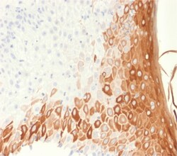
Biotium Cytokeratin 10 (Suprabasal Epithelial Marker) (rKRT10/1275), Biotin conjugate, 0.1mg/mL
This MAb recognizes a protein of 56.5 kDa, identified as cytokeratin 10 (CK10). CK10 is expressed in all suprabasal layers of the epidermis. In the epidermis, expression of CK10 strictly parallels the extent of differentiation; it is absent in the basal layer, appears in the first suprabasal layers and increases in concentration towards the granular layer. However, CK10 is rarely detected in early stages of vulvar squamous carcinomas (tumors less than 2 cm, clinical stage I) regardless of the tumor grade. In larger and more advanced tumors (greater than 2 cm, clinical stages II and III), CK10 is detected very frequently. Expression of CK10 is related to maturation of malignant keratinocytes, being preferentially detected in more-differentiated parts.Primary antibodies are available purified, or with a selection of fluorescent CF Dyes and other labels. CF Dyes offer exceptional brightness and photostability. Note: Conjugates of blue fluorescent dyes like CF405S and CF405M
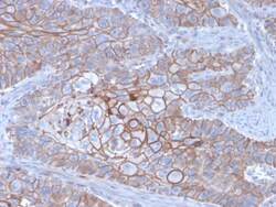
Biotium E-Cadherin (CDH1) / CD324 (Intercellular Junction Marker)(CDH1/3256), Biotin conjugate, 0.1mg/mL
Recognizes a protein of 120-80 kDa, identified as E-cadherin. Cadherins comprise a family of Ca2 -dependent adhesion molecules that function to mediate cell-cell binding critical to the maintenance of tissue structure and morphogenesis. The classical cadherins, E-, N- and P-cadherin, consist of large extracellular domains characterized by a series of five homologous NH2 terminal repeats. The relatively short intracellular domains interact with a variety of cytoplasmic proteins, such as -catenin, to regulate cadherin function. E-cadherin plays an important role in epithelial cell adhesion. A decreased expression of E-cadherin is associated with metastatic potential and poor prognosis in breast cancer, prostate and esophageal cancer. In combination with p120 Catenin, it is useful for the differentiation between ductal (E-cadherin ) and lobular (E-cadherin -) breast carcinomas. It may also help in diagnosis of mesothelioma. Primary antibodies are available purified, or with a s
Biotium CD44 Standard(DF1485), Biotin conjugate, 0.1mg/mL
Recognizes a cell surface glycoprotein of 80-95 kDa (CD44) on lymphocytes, monocytes, and granulocytes (Leucocyte Typing Workshop V). Its epitope is resistant to digestion by trypsin and chymotrypsin. The CD44 family of glycoproteins exists in a number of variant isoforms, the most common being the standard 85-95 kDa or hematopoietic variant (CD44s). Higher molecular weight isoforms are described in epithelial cells (CD44v), which are believed to function in intercellular adhesion and stromal binding. CD44 immunostaining is commonly used for the discrimination of urothelial transitional cell carcinoma in-situ from non-neoplastic changes in the urothelium.Primary antibodies are available purified, or with a selection of fluorescent CF Dyes and other labels. CF Dyes offer exceptional brightness and photostability. Note: Conjugates of blue fluorescent dyes like CF405S and CF405M are not recommended for detecting low abundance targets, because blue dyes have lower fluorescence
Biotium Nucleolin(364-5 + NCL/902), Biotin conjugate, 0.1mg/mL
Recognizes Nucleolin (NCL), the major nucleolar phosphoprotein of growing eukaryotic cells. NCL is located mainly in dense fibrillar regions of the nucleolus. It is found associated with intranucleolar chromatin and pre-ribosomal particles. Human NCL gene consists of 14 exons with 13 introns and spans approximately 11 kb. It has a predicted molecular weight of ~76 kDa with an apparent MW of 100-110 kDa, attributed to phosphorylation of its N-terminal domain. It induces chromatin decondensation by binding to histone H1. It is thought to play a role in pre-rRNA transcription and ribosome assembly.This MAb can be used to stain the nucleoli in cell or tissue preparations and can be used as a marker of the nucleoli in subcellular fractions. It produces a speckled pattern in the nuclei of cells of normal and malignant cells and may be used to stain the nucleoli of cells in fixed or frozen tissue sections. It can be used with paraformaldehyde fixed frozen tissue or cell preparations and
Biotium Beta-2-Microglobulin (B2M)(B2M/1118), CF568 conjugate, 0.1mg/mL
Beta-2 microglobulin is a component of the class I major histocompatibility complex (MHC I) where it functions in antigen processing and presentation. Involved in the presentation of peptide antigens to the immune system. Human Beta-2 microglobulin is expressed on many types of cells including lymphocytes, thymocytes, monocytes, granulocytes, platelets, endothelial cells, and epithelial cells. It is absent on erythrocytes. This MAb is specific to human Beta-2 microglobulin and does not react with non-human primate cells. This antibody reacts with all cell types excluding erythrocytes. The detection of Beta-2 microglobulin in body fluids has been used as a tumor marker and for monitoring patients with HIV infection.Primary antibodies are available purified, or with a selection of fluorescent CF Dyes and other labels. CF Dyes offer exceptional brightness and photostability. Note: Conjugates of blue fluorescent dyes like CF405S and CF405M are not recommended for detecting low ab
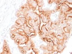
Biotium MUC16 / CA125 (Ovarian Carcinoma Marker)(MUC16/1860), 1mg/mL
The mucins are a family of highly glycosylated, secreted proteins with a basic structure consisting of a variable number of tandem repeats (VNTRs). Membrane-associated and secretory Mucins are high molecular weight glycoproteins of the glycocalyx (polysaccharide biofilm) that protects mucosal epithelium from particulate matter and microorganisms. Epithelial Mucins are large, secreted and cell surface glycoproteins crucial for adhesion modulation, signaling and epithelial cell protection. The number of repeats is highly polymorphic and varies among different alleles. The Mucin family consists of Mucins 1-4, Mucin 5 (AC and B), Mucins 6-8, Mucins 11-13 and Mucins 15-17. The Mucin 16 protein (also commonly referred to as CA125), encoded for by the gene MUC16, is a very high molecular weight tumor antigen consisting of three domains: a carboxy terminal domain, an extracellular domain and an amino terminal domain. Mucin 16, an ovarian cancer-associated antigen, is used as a marker
Biotium Epstein-Barr Virus (LMP-1)(CS4), 1mg/mL
This antibody is a mixture of four different monoclonal antibodies. This antibody is specific to 60 kDa latent membrane protein (LMP-1) encoded by the BNLF1 gene of the EBV. Each clone reacts with different epitopes on the hydrophilic C-terminus of the cytoplasmic domain of LMP-1. This antibody stains strongly with EBV-positive lymphoblastoid cell lines and EBV infected B cell immunoblasts in infectious mononucleosis. EBV, also designated human herpesvirus 4 (HHV-4), is a member of the herpesvirus family and is one of the most common human viruses. EBV infects B cells and, though often asymptomatic, it can cause infectious mononucleosis, a disease characterized by fatigue, fever, sore throat and muscle soreness. Primary antibodies are available purified, or with a selection of fluorescent CF Dyes and other labels. CF Dyes offer exceptional brightness and photostability. Note: Conjugates of blue fluorescent dyes like CF405S and CF405M are not recommended for detecting low
Biotium Galectin-1 / Human Placental Lactogen (hPL) (GAL1/1831), Biotin conjugate, 0.1mg/mL
Galectin-1 is a member of the beta-galactoside-binding family and is a dimeric protein of 14 kD participating in a variety of normal and pathological processes, including cancer progression. Galectin-1 can affect the proliferation of normal and malignant cells. Inhibition of cell growth is observed in a lactose-dependent manner as lower concentrations of the lectin stimulate cell proliferation. Galectin-1 may also be implicated in the induction of apoptosis of activated T cells through the binding of exogenous galectin-1 to CD45 molecules present on the surface of lymphocytes. Galectin-1, reported to be present either at the surface of cancer cells or accumulated around these cells could act as an immunological shield to protect against a T cell immune response and provide an advantage for survival.Primary antibodies are available purified, or with a selection of fluorescent CF Dyes and other labels. CF Dyes offer exceptional brightness and photostability. Note: Conjugates
Biotium Spectrin beta III (SPTBN2)(SPTBN2/1778), 1mg/mL
Spectrin is an actin binding protein that is a major component of the plasma membrane skeleton. Spectrins function as membrane organizers and stabilizers by forming dimers, tetramers and higher polymers. Vertebrate spectrins have two alpha-subunits (alpha-I/alpha-II), four beta-subunits (beta-I-beta-IV) and a beta-H subunit creating diversity and specialization of function. Spectrin α and spectrinβ are present in erythrocytes, whereas spectrin α II (also designated fodrin α) and spectrinβ I (also designated fodrinβ) are present in other somatic cells. The spectrin tetramers in erythrocytes act as barriers to lateral diffusion, but spectrin dimers seem to lack this function. Spectrinβ III is highly homologous to both spectrinβ I and spectrinβ II. Spectrinβ III is highly expressed in brain, kidney, pancreas and liver, and at lower levels in lung and placenta. Spectrin beta 3 is primarily expressed in nervous tissues with high